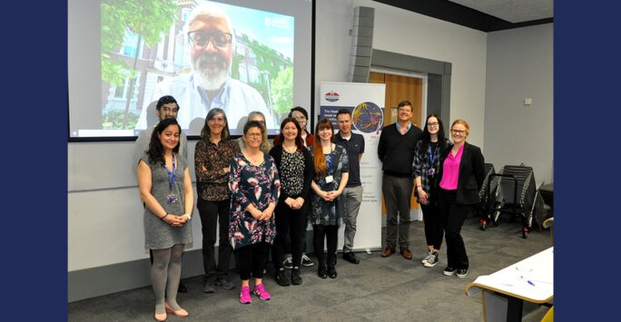
Over the summer, Dr Sharon Zytynska co-organised a workshop at the University of Liverpool, bringing together academic researchers and industry partners from the UK, Belgium and Canada. Here, she tells us all about the two-day workshop and her highlights.
I organised this workshop with Professor Joe Slupsky to explore ‘Developing imaging mass cytometry as a tool to study plant immune response at single-cell resolution’ and to welcome our international colleagues to Liverpool.
Imaging mass cytometry (IMC) is a technique that uses metal-tagged antibodies to visualise and measure multiple protein or mRNA targets in cells or tissue samples, allowing scientists to see the detailed makeup, structure, and function of tissues at a microscopic level.
Opening the workshop, we explored Imaging Cytometry as a tool with industry partners from Standard Biotools presenting the different options and upgrades for the instruments. This was followed by talks by Professor Dominique Van Der Straeten and Dr Olivier Leroux from Ghent University who were the first team to use Imaging Mass Cytometry on plant tissues.
Next up we learned about the set of data analysis pipelines being developed for IMC from Dr Bethany Hunter and Dr George Merces from the University of Newcastle.
To end this first session, Professor Joe Slupsky presented his novel approach for incorporating mRNA targets to IMC, using the Proximity Ligand Assay for RNA (PLAYR).
Exploring new research directions and techniques
The following session explored four research directions where IMC would be a beneficial tool:
- Plant cell-to-cell communication, Professor Robin Cameron (McMaster University)
- Root pathogen interactions, Professor Nat Kav (University of Alberta)
- Plant induced defences, Dr Sharon Zytynska (University of Liverpool)
- Plant stress responses, Professor Dominique Van Der Straeten (Ghent University)
The discussions after these talks highlighted many areas for collaboration and continued well into the evening when we met for dinner at a local restaurant, following a quick visit to the historic Philharmonic pub.
The next morning, we continued with a new set of presentations exploring alternative and integrative methods of cellular-level analysis of plants, the use of multiomics techniques for plant research with Dr Habibur Rahman (University of Alberta). We then took part in technical talks on the use of spatial transcriptomics (Dr Emily Johnson), metabolomics (Dr Howbeer Muhamed Ali), and insect imaging (Dr Marisa Merino).
After a final panel discussion summarising the current knowledge and technical gaps with ideas on how to develop these collaborations for future funding applications, we had a final lunch at the beautiful Waterhouse Café at the Victoria Gallery and Museum.
Building collaborations
The workshop was highly engaging and collaborative, offering insightful discussions into the use and potential uses of Imaging Mass Cytometry across our diverse research questions. The interactive format encouraged us to share ideas and strategies, fostering a dynamic exchange that highlighted new applications and inspired future collaborations.