Vision
The Reverend Dr. David C.M. Taylor
dcmt@liverpool.ac.uk
© David Taylor and The University of Liverpool, 1999
There is also a web page which gives a
simple overview of vision
These are notes from two lectures where we were specifically looking at receptor mechanisms. There is a whole world of visual physiology which I do not attempt to address.
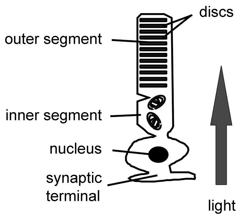
There are two types of receptors which are located between the retinal epithelium and the network of nerve cells and fibres; rods and cones.
Rods
 achromatic
slow response
long integration
single quanta
10 8
not in fovea
convergent pathways
.
Cones
 chromatic
fast response
short integration
low sensitivity
10 7
dense in fovea
divergent pathways
Transduction
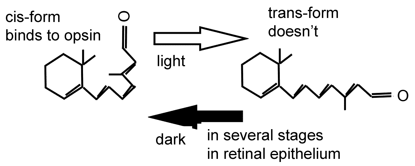
Chemical
Rhodopsin in rods is retinal + opsin
Cone opsin in cones (different forms of cone opsin absorb different wavelengths of light).
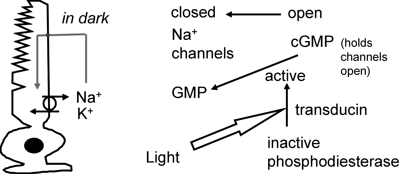
Electrical
Na+ channels are open in the dark
A single photon closes several hundred sodium channels, which results in a hyperpolarisation from -30 to -70 mV
This leads to a decrease in transmitter release.
The detail to remember (and try to work through) is called the dark current.
Retinal processing of information
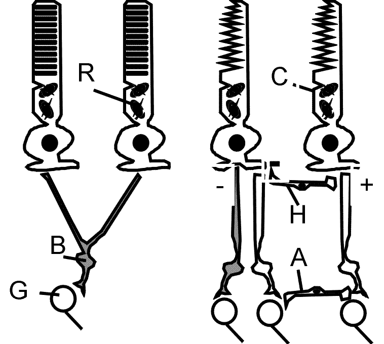
The retina contains a number of different types of cells, amongst them are
Rods - R
Cones - C
Bipolar cells - B
Horizontal cells - H
Amacrine cells - A
Ganglion cells - G
Intracellular recordings have allowed us to understand a considerable amount about the ways that retinal processing can occur. Remember that light shining on the retinal causes a hyperpolarisation of the membrane. If the transmitter released by the rod cell has excitatory effects, then we record an "off" response, and if the transmitter has inhibitory effects we record and "on" response.
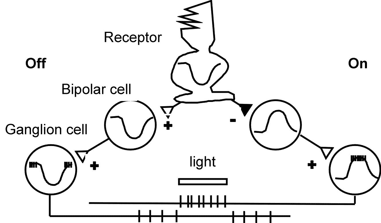
The situation is rather more complicated, however, because of the other nerve cells in the retina.
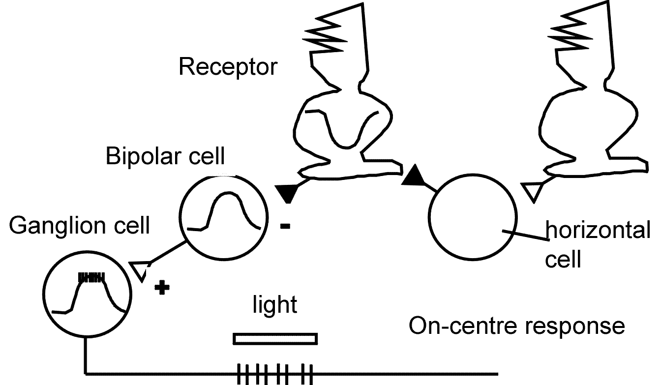
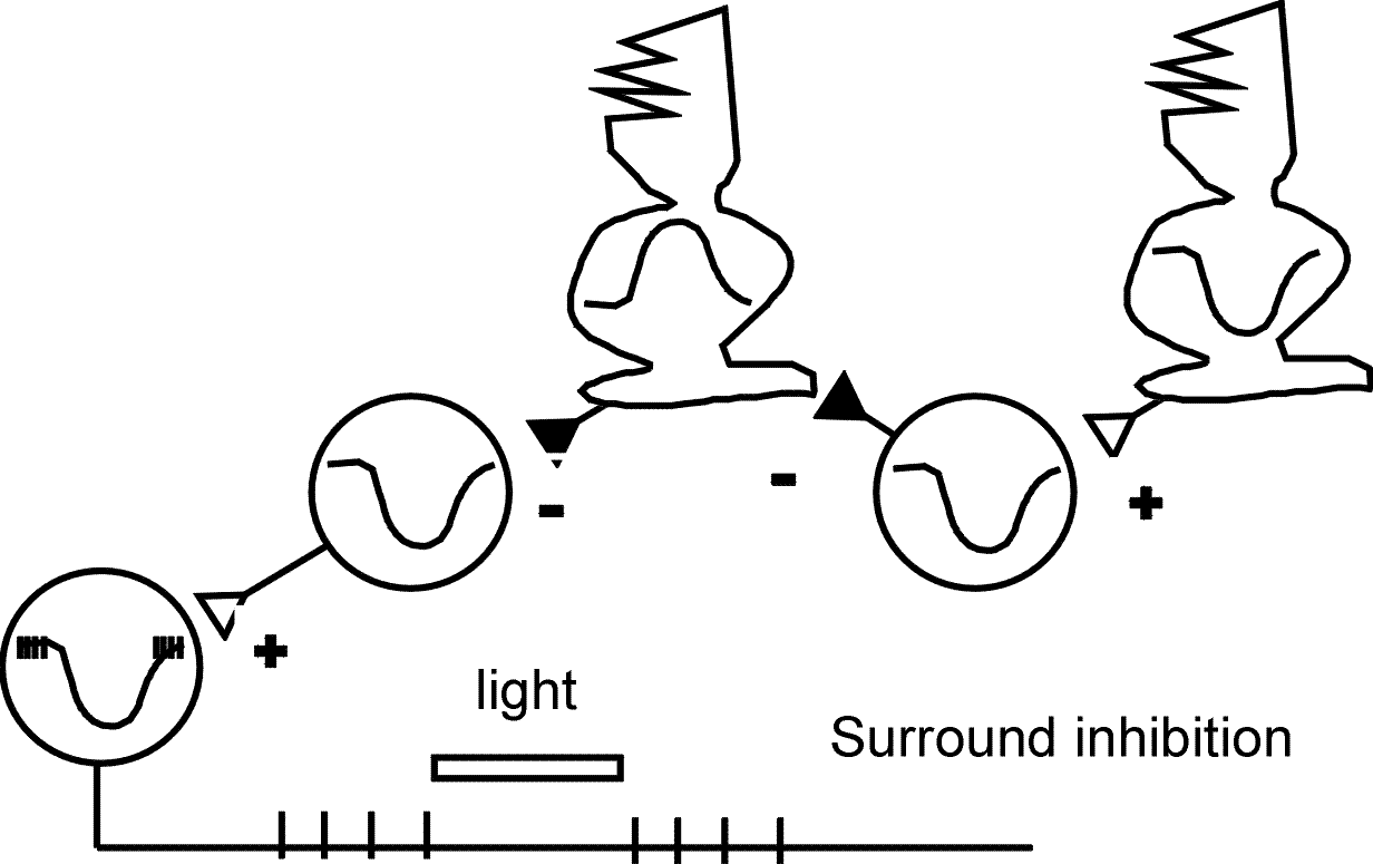
This is the conventional "on" response, but we know that surround inhibition is important in the retina, to provide contrast, and colour sensitivity.

back to top | back to index | back to simpler stuff