Spinal Reflexes
The Reverend Dr. David C.M. Taylor
dcmt@liverpool.ac.uk
© David Taylor and The University of Liverpool, 1999
 Chapter 5 in Carpenter, RHS, Neurophysiology 3rd Edition 1996, Arnold
Chapter 8 in Bray, Cragg, MacKnight, Mills & Taylor, Lecture Notes on Human Physiology 3rd Edition, 1994 Blackwell Scientific Publications
Chapter 37 in Kandel, Schwartz & Jessell, Principles of Neural Science 3rd Edition, 1991, Elsevier

Muscle sensors
Golgi tendon organs
Are arranged in series with the muscle fibres
Respond principally to changes in tension
are capsules within the tendons, 1mm long and 100m m in diameter
the intertwined nerve fibres are distorted during tension
Muscle spindles
Are arranged in parallel with the muscle fibres
Respond principally to changes in length of the muscles
There are 2 types:
nuclear bag fibres (dynamic response)
nuclear chain fibres (static response)
Each is innervated by a gamma motoneurone, which can therefore control muscle spindle length.
The consequence of the properties of the different muscle sensors is that we "know" when the muscle is either stretched or contracted.
|
Sensor |
Stretched |
Contracted |
|
Spindle |
+++ |
0 |
|
GTO |
+ |
+++ |
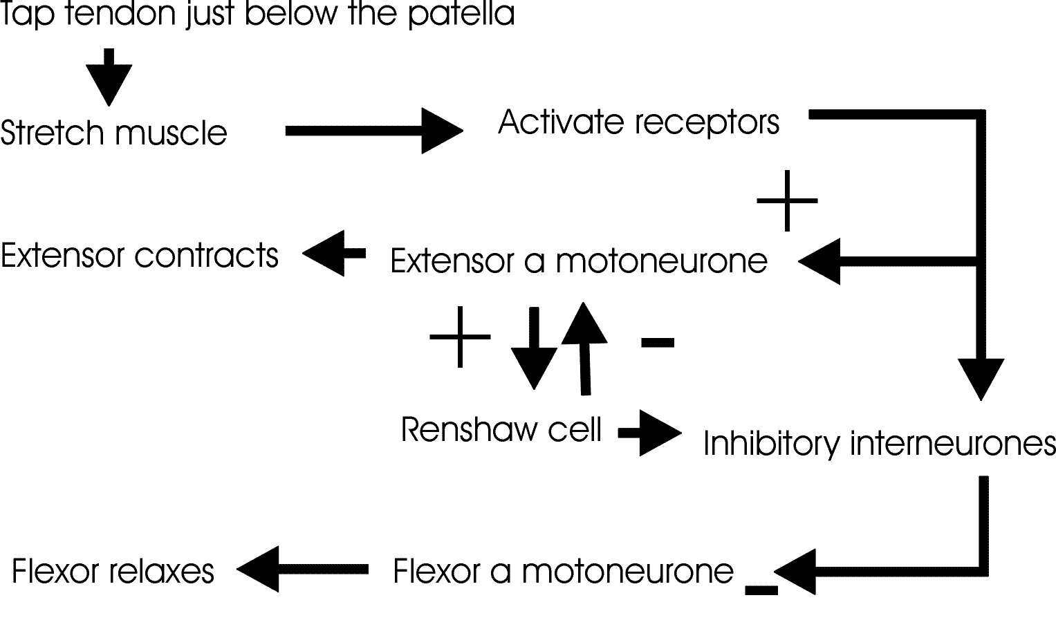
The monosynaptic reflex
A simple example shows the importance of the muscle spindle. The monosynaptic reflex is most easily observed as the knee jerk reflex.
Tapping the tendon just below the patella stretches the muscle, which includes the muscle spindles. These then cause the excitation of the extensor alpha motoneurones, and the inhibition of the flexor alpha motoneurons.. The reflex is turned off by the Renshaw cells, which are small interneurones innervated by co-laterals of the alpha motoneurones. The Renshaw cells provide an example of recurrent inhibition.

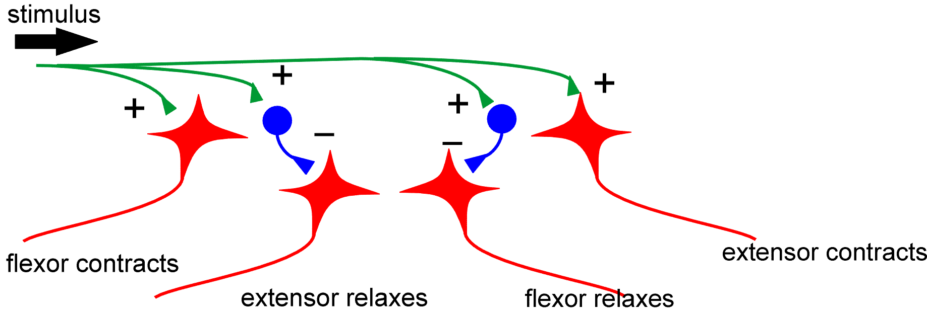
The Withdrawal Reflex
This is a simple reflex that hardly ever occurs in isolation (see below). When we reach out and (say) touch a hot object, we withdraw our hand. In other words it is a flexion reflex. In these terms, the knee jerk reflex is an extension reflex. In the withdrawal reflex, the flexor contracts, and the extensor is inhibited through an interneurone.

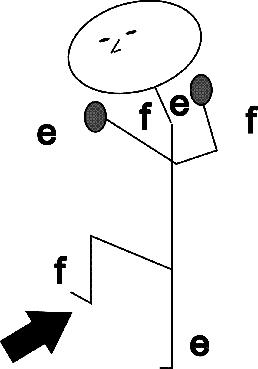
The crossed extensor reflex
The withdrawal reflex is, in reality, more complex than the reflex draw above, since a number of other reflexes occur, to allo us to maintain our balance. At its first level of complexity, the reflex is as shown below, with flexion on the ipsilateral side (the same side as the stimulus) and extension on the contralateral (opposite) side - hence the name "crossed extensor reflex".
The withdrawal reflex is normally only observed as a crossed extensor reflex. But, remember that the crossed extensor reflex occurs at each hierarchical level of the nervous system. This explains quite a lot of the complex responses that occur in response to a sharp, short lasting stimuli.
back to top | back to index