The electrocardiogram (ECG).
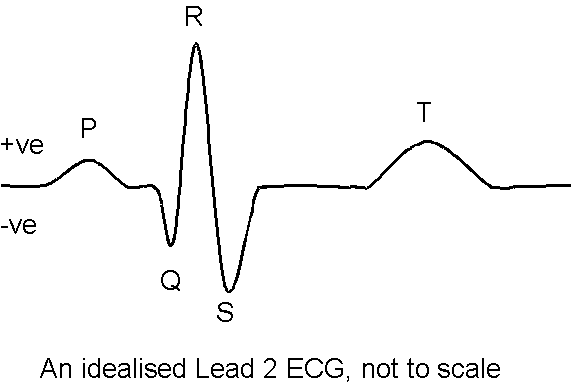
The electrocardiogram is covered in nearly all physiology text-books, and in 2LS28 you will only be expected to be able to interpret a normal Lead 2 ECG, explain what each of the waves represents and have some concept of what the ECG can be used for.
The ECG is a representation of the electrical activity of the heart, which we record from electrodes attached to the body surface. The electrical signal is quite small, and interference can be a problem, so we usually attach one of the electrodes to act as a reference and another electrode to "ground", to minimise the effects of electrical interference.
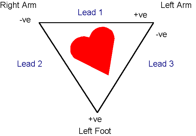
There are an infinite number of ways that electrodes could be placed on the body to record the electrical activity of the heart, so a convention has been drawn up, which allows the results from one laboratory to be compared with those from any other laboratory in the world. The convention which we shall use is based on Einthovenís triangle. This uses bipolar leads (one negative and one positive), and by convention any wave of depolarisation passing towards the foot is recorded as a positive deflection, and any wave of depolarisation passing towards the right shoulder is recorded as a negative deflection.
Einthovenís Triangle
Excitation normally starts in the sinoatrial node (SAN), and passes over and through the walls of the atria. This is represented on the ECG by the P-wave, and corresponds to the period of atrial contraction.
The waves of excitation then pass through to the atrioventricular (AV) node and spread through the Bundle of His, which is composed of Purkinje fibres. The excitation passes first into the intraventricular septum, and the direction of spread of current through the intraventricular septum is towards the right shoulder (Q-wave).
The main spread of current through the ventricles, however is towards the foot, and this is represented by the large positive R-wave.
Excitation of the ventricles is completed by the S-wave, as the excitation spreads around to an area of the ventricles at the "back" of the heart.
Repolarisation occurs in the reverse order, and is observed as a positive T-wave. Most people need to think about this quite carefully before they finally understand why!

This page © David Taylor/The University of Liverpool 1999