X-ray Computed Tomography Imaging of Decellularised Human Trabecular Meshwork
This study investigated the potential to use of x-ray computed tomography to observe the decellularised structure of the human trabecular meshwork, a minute, porous tissue found in the iridocorneal angle of the eye and known to be involved in the progression of glaucoma.
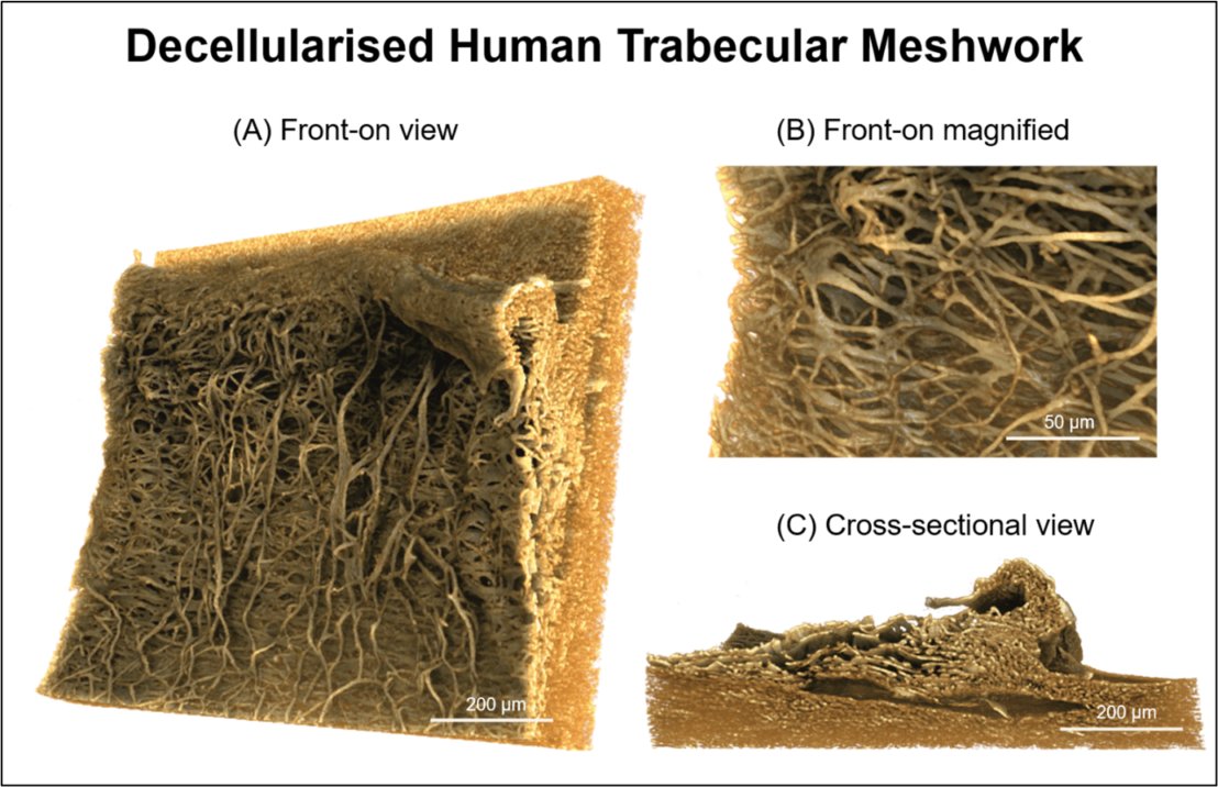
Glaucoma is the 2nd leading cause for irreversible blindness worldwide and is linked to raised intraocular pressure (IOP). The trabecular meshwork (TM) plays a major role in maintaining homeostatic IOP by regulating outflow of aqueous humour from the eye through its intricate 3D structure. A lack of therapies targeting diseased TM highlights the need to develop biomimetic scaffolds that provide 3D in vitro models for glaucoma research or as implantable devices to regenerate TM tissue. Before artificially replicating the TM’s structure, we first need to visualise the tissue’s architecture in 3D. However, cells act as obstructions to the TM’s structure and hinder our ability to observe the intricacies of the tissue. Therefore, to mimic the TM, we first optimised a decellularisation method for cell removal and structural retention. Decellularisation will permit visualisation of the TM void of cells to truly appreciate the complexity of the tissues structure. X-ray computed tomography enabled a novel 3D reconstruction of decellularised human TM and observation of the porous tissue’s intricate architecture.
More details about the decellularisation protocol can be found in the Open Access article An Optimized Method to Decellularize Human Trabecular Meshwork
This data has also been used for modelling the aqueous outflow pathway as published in Science Direct.

Images and videos shown here were created with Drishti from Zeiss Xradia Versa data.