X-ray CT Research Example: Dissolvable Microneedle Array Patches
Dissolvable microneedle array patches (DMAPs) combine characteristic advantages of injected therapeutics, such as efficient dosing, whilst retaining a minimally invasive application similar to topical administration (TA) due to the sub-micron size of the needles.
This project “Degradable microneedle arrays for user-directed nanomedicine delivery for the treatment of glaucoma” involves the development of DMAPs for ocular administration. Glaucoma is the 2nd world leading cause of irreversible blindness. It is associated with a rise in intraocular pressure (IOP) linked to inadequate drainage of aqueous humour through the trabecular meshwork (TM). All treatment methods for glaucoma aim to reduce the IOP via increased outflow or decreased production of aqueous humour from the ciliary body. Current treatments include the TA of active pharmaceutical ingredients; however, this is heavily limited by poor permeability and retention, or highly invasive surgical procedures. A solution to combat issues surrounding corneal drug administration is the use of DMAPs.
DMAPs are fabricated using water soluble materials, such as polymers, whereby the therapeutic is encapsulated within the polymeric matrix. Upon contact with a hydrophilic environment the polymer undergoes dissolution, and allows the targeted delivery of the therapeutic.
X-ray microcomputed tomography has enabled the architecture and topography of the DMAPs to be visually observed and analysed. This technique has allowed for the effects of polymeric composition on needle structure to be determined. Subsequent physiochemical analysis aims to target DMAPs which offer fast dissolution whilst maintaining structural integrity to maximise successful permeation into tissue with minimal breakages and deformation.
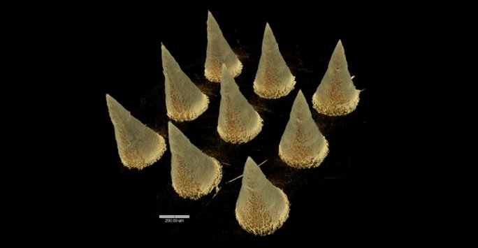
The X-ray microtomographic images below show a bespoke 3x3 DMAP. Scale bar = 200 µm.
For more details about this project, contact Elliot Croft or Dr Helen Cauldbeck.
The video and images shown here were created with Drishti from Zeiss Xradia Versa data.