Micro XCT research example: Analysis of 3D printed gelatin scaffolds
Micro X-ray CT imaging can be used to investigate pore size, wall thickness, and connectivity of porous structures, such as 3D printed gelatin scaffolds for dental applications.
Example of pore size and wall thickness analysis of gelatin scaffolds
X-ray Computed Tomography Imaging of 3D printed Gelatin Scaffolds loaded with Quaternary Ammonium Compounds for dental applications.
Regenerative Endodontic Procedures (REPs) are dental treatments for immature permanent teeth that aim to restore tooth health and function. Recent research has shown that up to 79% of failed REPS are due to persistent bacterial infections. The use of antimicrobial materials that can reduce bacterial burden, whilst maintaining regenerative function, would reduce the failure rate of these procedures. The aim of this research is to develop 3D printed scaffolds loaded with antimicrobial compounds that can ensure sufficient antibacterial efficacy and create a favourable environment for cells to regenerate the lost or damaged structures of the tooth. The 3D structure and porosity of the scaffold has an important role in drug release, facilitating cell infiltration, proliferation, and guiding scaffold degradation and tissue remodelling processes. Micro-CT imaging has been key to the development of this work as it has produced essential information in relation to the porosity and interconnectivity of quaternary ammonium compound loaded 3D printed gelatin scaffolds.
For further information about this project, please see the publication: 3D printable gelatin/nisin biomaterial inks for antimicrobial tissue engineering applications.
Another recent application describes the use of the scaffolds: Antimicrobial 3D printed gelatin scaffolds for root canal disinfection in regenerative endodontics procedures.
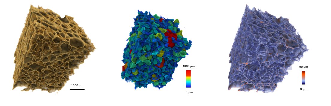
The video and images shown here were created with Fiji and Drishti from Zeiss Xradia Versa data.