High penetration 3D/4D imaging
High penetration of samples can be achieved on the light sheet microscope where the fluorescence light is shaped as a thin sheet and used to excite a single plane inside the sample.
The emitted fluorescence light is captured perpendicularly using a camera making the technique fast, gentle and suitable for large specimen.

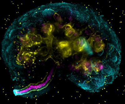
Mouse embryonic kidney stained for basement membrane (cyan (limited penetration) & yellow (full penetration)) highlighting the ureteric tree and the ureteric lumen (magenta) which make up the tubes leading the filtered urine to the bladder.
Another high penetration technique available in the CCI is multiphoton imaging. It uses near-infrared excitation light, resulting in suppressed background signal and increased depth penetration. This equipment also provides second harmonic generation microscopy.

Second harmonic generation signal of collagen fibres. The image was recorded as a z-stack and the different heights were colour coded (bottom – black, middle – cyan, top – white). Data courtesy of Jill Madine and Riaz Akhtar labs
Microscopes: