New advancements in tumour imaging
Former OMA fellow Navrit Bal, now a postdoc at the Aarhus University Hospital in Denmark, is part of a team developing a spectral photon-counting micro-CT system.
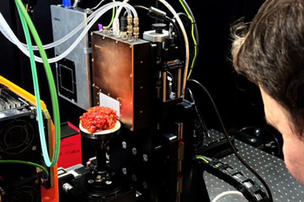
The device, based on a Medipix3 detector, can obtain the spectrum of the x-rays that pass through a tumour. This spectral information is expected to be different between tumour and surrounding normal tissues, providing more imaging contrast. By determining the tumour-normal tissue boundary during surgeries, re-operations can be reduced, improving survival rates.
The team led by Jasper Nijkamp has recently reached a milestone, coinciding with World Cancer Day, by scanning their first breast tumour surgery specimen with the spectral photon-counting micro-CT system. This marks a significant step toward online surgical margin assessment, with one clear goal: reducing re-operations for breast cancer patients.
Navrit says: “We’ve been working towards this quite literally for years now – it’s very satisfying to finally start this phase of pre-clinical research!”
Feature image: PhD candidate Danny Mortensen, starts his project with the first tumour scan from a breast cancer patient at Aarhus University Hospital.