Dr Ahmed Abass PhD, CPS, MSc, BEng (Honours, 1st class), FHEA, CEng MIMechE
Lecturer in Biomedical Engineering Materials, Design and Manufacturing Eng
- Work email A.Abass@liverpool.ac.uk
- Personal WebsiteResearchGate
Research
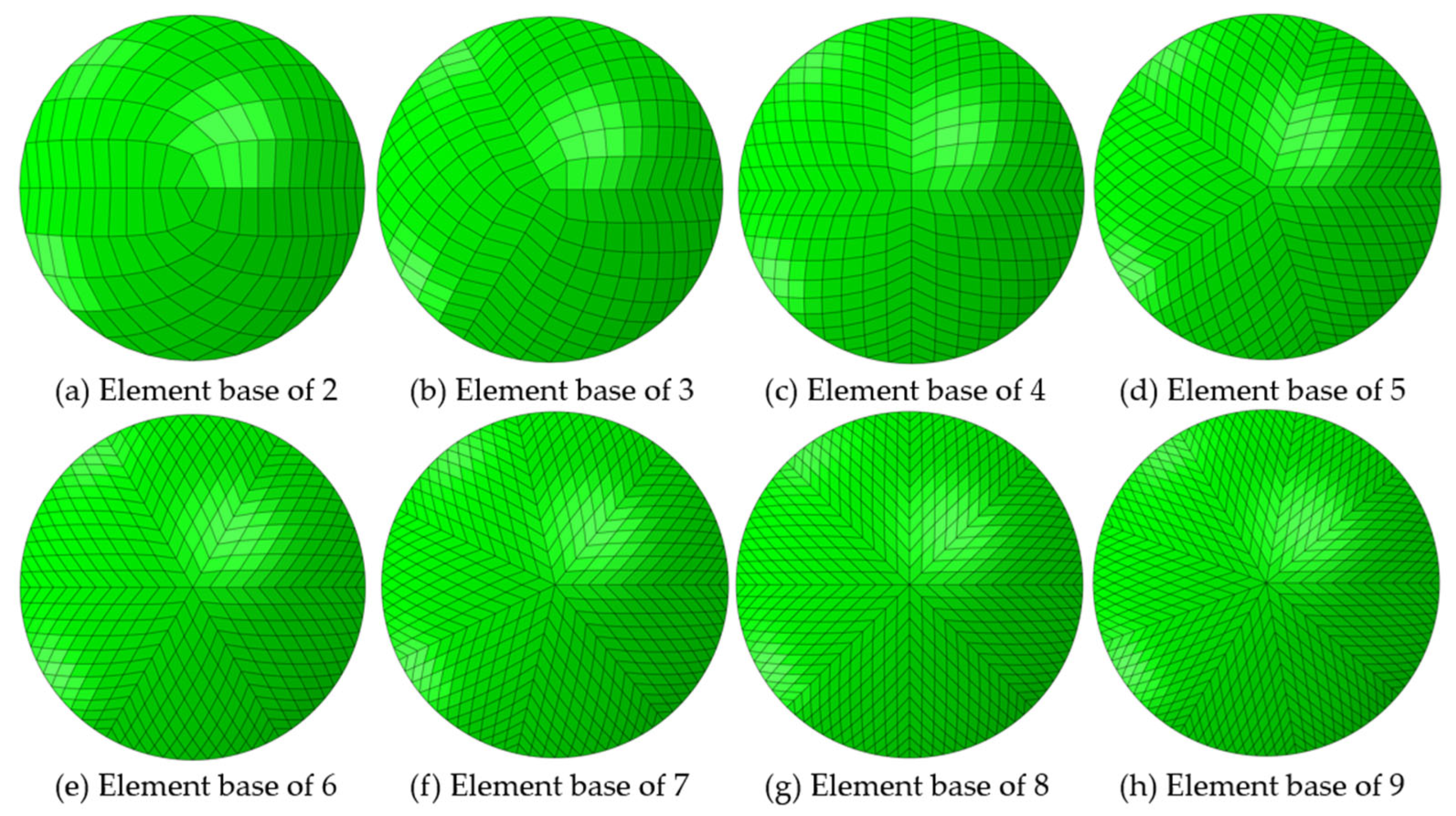
Nonparametric meshing algorithms

Nonparametric meshing algorithms
Creating non-parametric meshing algorithms allows the meshing of both regular and irregular geometries with uniform meshes. The algorithm was built to model contact lenses, but it was recently updated to be used in various applications. Click here to see a graphical representation
Abass, A., et al., Simulated optical performance of soft contact lenses on the eye. PLOS ONE, 2019. 14(5): p. e0216484.
Doll, T., et al., Which feature influences on-eye power change of soft toric contact lenses: Design or corneal shape? PLOS ONE, 2020. 15(11): p. e0242243.
Shihab, A.H., et al., Compressive behaviour of soft contact lenses and its effect on refractive power on the eye and handling off the eye. PLOS ONE, 2021. 16(2): p. e0247194.
Towler, J., et al., Typical localised element-specific finite element anterior eye model. Heliyon, 2023. 9(4).
If you are a student looking for a PhD opportunity, you may like to have a look at the following PhD projects: Digital soft contact lenses design and validation and Innovating an anti-tilt human eye topographer.
Microstructure of ocular tissues

Dr Abass investigated the micro-structure of corneal collagen distribution and orientation over the corneal cross-section. Following the suggestion that corneal surface topography may be stabilised by the angular orientation of out-of-plane lamella that insert into the limiting membrane of the anterior cornea, his research aimed to provide quantitative information about the angular orientations of these lamella across and throughout the depth of the cornea. Wide angle x-ray scattering experiments were carried out on beam-line IO2 at the UK’s national synchrotron facility, Diamond Light Source (Oxfordshire, UK).
If you are a student looking for a PhD opportunity, you may like to have a look at the following PhD projects: Digital soft contact lenses design and validation and Innovating an anti-tilt human eye topographer.
Contact lenses modelling

Dr Abass's project with UltraVision has been graded by a panel of independent assessors as Grade A (Outstanding).
The project developed state-of-the-art contact lenses for people with vision loss due to irregular corneas. A suite of software codes was built to deal with all types of distorted corneas. This now allows any corneal profile that can be measured by an eye topography device to be modelled in our advanced FE software. The original objective in the project was to prepare for the second generation of the company KeraSoft product, however, the outputs obtained were designed to be flexible enough to be used for developing all the contact lenses produced by the company. This included the new generation of the KeraSoft lens and the recent product, Avanti (a monthly disposable contact lens).
If you are a student looking for a PhD opportunity, you may like to have a look at the following PhD projects: Digital soft contact lenses design and validation and Innovating an anti-tilt human eye topographer.
Research Grants
Rami Rasheed Alanazi - Bench fees (201606338)
ROYAL EMBASSY OF SAUDI ARABIA CULTURAL BUREAU IN LONDON (UK)
May 2023 - April 2027
Bench fees EMAN YOUSEF A AL AHMAD - (201711883)
ROYAL EMBASSY OF SAUDI ARABIA CULTURAL BUREAU IN LONDON (UK)
June 2023 - May 2027
Development of soft orthokeratology lenses
SEED (JAPAN)
November 2020 - November 2028
Spectacles for correcting irregular astigmatism in keratoconic patients
FIGHT FOR SIGHT (UK)
January 2021 - December 2022
Research Collaborations
Prof Jos Rozema
Project: Tilted eye optics
External: University of Antwerp, Belgium
Optical axis analysis.
Dr Alejandra Consejo
Project: Eye geometry analyses
External: University of Zaragoza, Zaragoza, Spain
Eye topography.
Prof Renato Ambrósio Jr
Project: Corneal topography
External: Federal University of São Paulo, São Paulo, Brazil
New KC indices
Dr Craig Boote
Project: Micro-structure of ocular tissue
External: Cardiff University, Cardiff, UK
The use of X-ray to identify the micro-structure of ocular tissue