This new drug delivery method could offer long-lasting relief for eye diseases
University of Toronto Engineering researchers are working to enable a future where a single, non-invasive injection under the eyelid could replace months of daily eye drops in treating glaucoma, a leading cause of blindness.
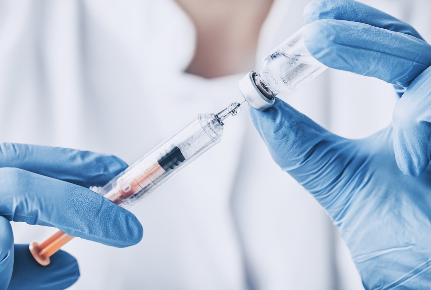
In a recently published paper in Advanced Materials, a team led by Professor Molly Shoichet (ChemE, BME) describes how they used colloidal drug aggregates (CDAs) to modify the effects of a small-molecule glaucoma drug. This new approach prolongs the drug’s effect from six hours when it is delivered via an eye drop, to up to seven weeks with a single, non-invasive injection placed in the subconjunctival space, that is, under the eye lid.
Glaucoma is a group of eye diseases characterized by an increase in eye pressure, leading to damage of the optic nerve, which is essential for vision. Currently, there are no clinical cures, only treatments that can slow the progression of the disease.
Eye drops are the most common treatment for glaucoma, but they come with issues regarding efficacy and patient compliance, especially since the disease is more common in older adults. Self-administering drops perfectly can be difficult, and their effects are transient, requiring administration on a precise, interval-based schedule. There are also laser therapies and surgical treatments that require an injection inside the eye every few months. But these come with risks of complications, such as infection, inflammation or vision loss.
says Mickaël Dang (ChemE PhD 2T4), postdoctoral fellow in Shoichet’s lab and the first author of the study.
The new method delivers timolol prodrug colloids dispersed in a hydrogel, demonstrating for the first time that a non-colloid forming drug can be chemically modified into a colloid-forming prodrug.
CDAs are drug molecules that can spontaneously self-assemble into nano-scale particles. Traditionally, they have been seen as a hindrance in drug development research. This is due to CDAs creating false positive and false negative results in enzyme- and cell-based assays, respectively, which are commonly used to screen and characterize drug candidates in the early stages of development.
We showed that delivery of this colloidal drug aggregate could be dispersed in an in situ forming hydrogel into the subconjunctival space. The colloidal drug enabled the slow release over several weeks leading to a 200-fold increase in efficacy, and the hydrogel resulted in the formulation staying in the subconjunctival space after injection. The control without the hydrogel mostly leaked out of that space.
says Shoichet.
Shoichet’s lab collaborated with Dr. Jeremy Sivak on this research. Sivak is an associate professor in Temerty Faculty of Medicine’s Department of Ophthalmology & Vision Science and the glaucoma research chair at the Krembil Research Institute, part of the University Health Network.
While this study was conducted on animal models, the researchers are now working towards optimizing their formulation for ultimate clinical use.
We envision a future where this non-invasive injection can be administered once every month or two in a medical office. We invented this novel hydrogel as a vitreous substitute for vitreoretinal surgery, and here we show its versatility to encapsulate and release small molecule drugs.
says Dang, who is also at Synakis, a spinoff biotechnology company founded from research in Shoichet’s lab.
“There is a lot of work ahead,” adds Shoichet. “We are focused on the stability and manufacturability of our product while at the same time looking to raise funds to advance it more quickly to the clinic.”
For more information, read the original press release.
For more news from the world of long-acting therapeutics, sign up to the CELT's LONGEVITY mailing list here for regular updates.